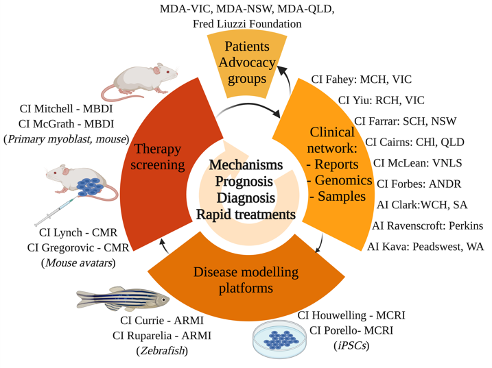
Our research
For decades, congenital muscle diseases have remained an intractable health issue due to the lack of treatments. The major hurdle remains understanding the underlying molecular basis of disease, coupled with a lack of patient specific cell- or animal-based disease models to develop and test therapies.
The Genomic Revolution has led to major advances in diagnosing congenital muscular diseases however, this genomic diagnosis frequently falls short in providing clinically meaningful answers to:
- Accurate predictions of how a disease will progress in individuals with specific mutations
- Innovative therapeutic interventions that can be fast tracked into clinical trials
- Identifying biomarkers, to detect disease occurrence, progression and for use in clinical trial evaluations
By assembling a national network including paediatric neurologists and scientists we will leverage this world class expertise to find new treatments to better understand disease mechanisms and test therapies that can be fast-tracked into clinical trials.
Using various research models - fish, mouse and patient-derived cells - we will be able to provide new functional diagnostic tools, improve prognostic measures and test new drugs in the laboratory.
-
Zebrafish Models
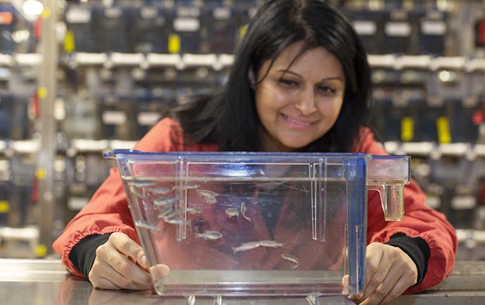 We have a long-standing interest and success in modelling muscle diseases using zebrafish, which given their prolific reproduction, amenability to genetic manipulation, optical transparency to enable live imaging, and high degree of conservation in muscle structure and function to humans, make them optimal for muscle disease research. We will leverage on these unique advantages of the zebrafish model to identify disease mechanisms and therapies for congenital muscular dystrophies.
Using CRISPR/Cas9 knockout and knock-in strategies, and transgenesis techniques, we will generate novel models of congenital muscular dystrophies. We will subsequently use a combination of immunofluorescence, western blot experiments, live imaging, -omics based approaches, and muscle function assays, to examine muscle structure and pathology of our newly established models. These results will reveal the cellular and molecular mechanisms responsible for the pathogenesis of muscle wasting in congenital muscular dystrophies, leading to the development of meaningful clinical outcomes such as diagnostic and prognostic biomarkers for evaluating disease progression and clinical trial outcomes.
Having identified disease mechanisms, we will utilise our successful drug screening pipelines to identify rapidly translatable therapies that not only improve muscle pathology but also muscle function for the treatment of congenital muscular dystrophies.
Dr Avnika Ruparelia
Centre for Muscle Research, Department of Anatomy and Physiology, The University of Melbourne
Professor Peter Currie
Australian Regenerative Medicine Institute, Monash University
Contact
Dr Avnika Ruparelia
Email - Avnika.ruparelia@monash.edu
Mouse Models
 Mice are one of the most widely used model systems to study congenital muscle disease, and are invaluable for understanding disease mechanisms. Pre-clinical trials in mouse models that evaluate novel therapies can also provide critical proof of concept knowledge prior to scaling into human clinical trials. These mouse models are generated via genetic modification, using a range of approaches including the Cre-LoxP- or CRISPR-gene editing tools, to introduce mutations that cause muscle disease in patients. Enabled by our state of the art metabolic phenotyping platform at the Monash Biomedicine Discovery Institute, we have a suite of specialised instrumentation to monitor muscle disease progression in mouse models, . Our approaches are analogous to patient examination methodologies including EchoMRI to image muscle mass, muscle (grip) strength measurement, real-time monitoring of activity and exercise tolerance (treadmill running), muscle metabolism, and serum profiling including measurement of serum creatine kinase as a measure of muscle disease. We are also uniquely positioned to integrate this with some of the most advanced microscopy techniques available such as live-cell and super-resolution microscopy and volumetric 3D-electron microscopy, including of patient-derived cells,. This in-depth knowledge has the capacity to identify functionally characterise new therapeutic targets. We have a particular interest in diseases linked to alteration of phosphoinositide and autophagy pathways, and also those caused by mutations in the FHL1 gene.
Professor Christina Mitchell
Dr Meagan McGrath
Monash Biomedicine Discovery Institute, Monash University
Contact
Dr Meagan McGrath
Email - meagan.mcgrath@monash.edu
Patient derived Stem Cell Models
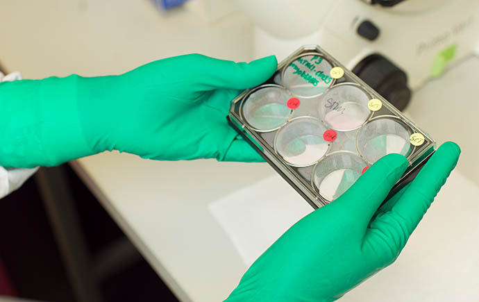 Using peripheral blood mononuclear cells from patients obtained by the clinical network team, we will generate iPSC lines and matched isogenic controls, a process which is well established within our team as we have already generated many novel iPSC lines. We will generate up to 3 patient specific iPSC lines per year (with corrected isogenic controls) across the 5-year project duration. Following iPSC generation and validation we will differentiate these cells to myotubes (imyotubes) to examine disease and patient specific phenotypes (including autophagy and Golgi disruption). Additionally, differentiated iPSCs cells will be grown as 3D skeletal muscle bundles with advanced maturation properties, for use in assessing muscle force and adhesion using our established methods, to define patient-specific effects on muscle function. If effects on our nominated pathways are detected, we will then progress into the therapy development pipeline.
Transplanting human iPSCs into animal models
To effectively study the patient’s disease, iPSC’s and their derivatives may be grown in other species (such as mice) to provide a proper setting for grow and development. These preclinical model systems will then be used to discover how muscle disease develops and be used to identify and test potential new treatments. This will be performed in collaboration with Prof Gordon Lynch and Paul Gregorevic.
Professor Enzo Porrello
Dr Peter Houweling
Murdoch Children's Research Institute
Professor Catriona McLean
Anatomical Pathology , The Alfred
Contact
Email - Pete.houweling@mcrie.edu.au
Mouse Avatars Models
Information to follow.
Professor Gordon Lynch
Muscle Research and Therapeutics, Department of Anatomy and Physiology, University of Melbourne