Protein structure


Use this page to revise the following concepts within protein structure:


Protein structure

Medications often target enzymes to regulate biochemical processes in the body. Understanding the structure of proteins is a key to understanding enzyme function, as almost all enzymes are proteins. This knowledge enables scientists to design medications that precisely target enzymes for desired therapeutic effects.

2-amino acids

Amino acids are the building blocks of all proteins. There are twenty different amino acids found in living things. They have in common an amino group and a carboxyl group attached to a central carbon atom. They differ by the R group attached to this central carbon atom.

As the name suggests, 2-amino acids contain an amino group, attached to carbon 2, and a carboxyl group, bonded to a common carbon atom.

A visual representation of the amino acid structural formula. To the left of the image is a vertical rectangle with the letters H N H, connected to each other via lines, in the centre. This rectangle is labeled amino group. To the right of this rectangle, connected via a line, are the letters HCR vertically arranged with lines connecting each letter to its neighbour. This group of letters connects, via a line, to another rectangle on the right side of the image. In this rectangle are the letters O, connected via two parallel lines to the letter C, which is connected via one line to the letters OH. This rectangle is labeled carboxyl group.

There are twenty amino acids that differ only by the R groups attached to the central carbon atom. Three examples are shown below, with the R group circled in blue.

An image of the chemical structure of 3 amino acids. The left-hand side amino acid is labeled glycine and a dotted circle encompasses part of the structure depicted by a H with a vertical line above it. The centre amino acid is labeled alanine and a dotted circle encompasses part of the structure depicted by the letter C in the centre of the circle with 4 lines extending from it at equal points around. 3 of these points connect to a letter H and the final line extends back toward the rest of the structure. The third amino acid, on the right side is labeled serine and a dotted circle encompasses part of its structure depicted by the letter C in the centre of the circle with 4 lines extending from it at equal points around. The lines to left and right of the circle point to a letter H, the line descending vertically from the circle points to the letters OH and the line ascending vertically from the circle connects back with the rest of the amino acid structure.

The nature of the R group determines the solubility of the amino acid. Alanine, with a non-polar methyl group, has a lower solubility in water than serine, with a polar hydroxyl group.

The R groups on many amino acids are larger and more complex than those above, as shown in the examples below:

A graphic depicting the chemical structure of lysine, glutamine and tryptophan.

Zwitterions

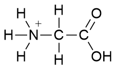
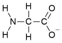
Amino acids can exist in aqueous solutions as a dipolar ion, known as a zwitterion. This ion forms as the carboxyl group donates a proton to the amino group on the other end of the molecule. The diagram below shows glycine and its zwitterion. Zwitterions are always electrically neutral, as their charges cancel out.

An image with the chemical structure of glycine depicted on the left side and the chemical structure of the glycine zwitterion on the right side.

The pH of different organs and regions of the body varies. The overall charge of an amino acid depends upon the pH of the surrounding environment. For example,

  • Glycine will form a cation in acidic conditions (low pH) due to the presence of H+ ions.

The chemical structure of glycine zwitterion with charged amine.

  • Glycine will form an anion in alkaline conditions (high pH) due to the presences of OH- ions.

The chemical structure of glycine zwitterion with charged carboxyl.

Levels of protein structure

Proteins are large molecules formed from condensation reactions between amino acids. They have complex shapes that allow them to facilitate various reactions in living things. An understanding of this shape involves learning about the levels of structure of protein. These levels are labelled primary, secondary, tertiary and quaternary.

The carboxyl group on one amino acid bonds readily to the amino group on another amino acid. The products are a dipeptide and water. The bond joining the amino acids is called a peptide link or an amide link. The formation of a dipeptide is an example of a condensation reaction, as water is also formed.

A graphic depicting amino acid condensation to dipeptide. On the left hand side of the graphic the chemical structure for alanine is depicted. To the right of this image is a plus sign. To the right of the plus sign is an image of the chemical structure for glycine. To the right of this image is an arrow pointing to the right. To the right of the arrow is a chemical structure labeled dipetide: alanine-glycine. To the right of this chemical structure is a plus sign. To the right of this plus sign is the chemical structure for water, H2O.

More amino acids can bond to the carboxyl end and amino end of the dipeptide via further condensation reactions and eventually form a polymer.

A graphic depicting amino acid condensation to tripeptide. On the left hand side of the graphic a chemical structure is depicted labeled dipetide: alanine-glycine. To the right of this image is a plus sign. To the right of the plus sign is an image of a chemical structure labeled alanine. To the right of this image is an arrow pointing to the right. To the right of the arrow is a chemical structure labeled tripetide: alanine-glycine-alanine. To the right of this chemical structure is a plus sign. To the right of this plus sign is the chemical structure for water, H2O.

Proteins can have thousands of amino acids in a long polypeptide chain. Insulin, a hormone that regulates blood sugar levels, containing 51 amino acids, is one of the smaller proteins.

However, proteins are not simply long, linear molecules. They have several structural features that lead them to fold into intricate shapes that allow the biological functions in a living organism.

Primary structure

The sequence of amino acids in the protein is referred to as its primary structure. Shorthand notation using the three letter abbreviation for each amino acid is often used to show the primary structure.

 A graphic depicting a primary structure. A curved line comprising small circles with the edges touching. Two arrows point at two of these small circles. The two arrows converge on a label that reads amino acid subunits. One end of the curved line is labeled H3N amino end.

Secondary structure

The presence of oxygen and nitrogen atoms in protein chains leads to significant dipoles on the peptide links (see the diagram below).

The chemical structure of a partial peptide with partial charges.

These dipoles enable hydrogen bonding between the polypeptide backbone of different parts of the same protein chain, which drives the formation of the protein's secondary structure. This bonding pulls the protein into specific shapes, such as a helical structure or a pleated sheet as shown below.

A graphic labeled secondary structure. On the right hand side of the graphic is an image depicting a protein secondary structure pulled into a helical shape. On the right hand side of the graphic is an image depicting a protein secondary structure pulled into a pleated sheet shape.

Tertiary and quaternary structures

Interactions between the R groups on amino acids lead to further shaping of the protein chains and gives each protein its unique 3D structure. The diagram below highlights various types of bonds that can form between R groups. The bonding types include:

  • covalent disulfide bond (between R groups from two cysteine amino acids)
  • dispersion forces (between non-polar R groups)
  • hydrogen bonding (between -OH, -COOH or -NH groups within R groups)
  • ionic bonds (between COO-formed from carboxyl groups and NH3+ formed from amino groups).

A graphic depicting tertiary quaternary structures. The graphic consists of a curving blue line that flows from the left side of the graphic to the right side. On the left side the blue line is curved into a spiral, and this section is labeled alpha-helix. Between the spiral and a curve of the blue line to the right is a chemical structure labeled dispersion forces. To the right of this the space between two curves of the blue line are a series of dotted parallel lines, connecting one side of the curve to the other. At the bottom of this curve, as the blue line curves back up, there is another chemical structure depicted. The upward curving blue line, at this stage, is labeled beta-pleated sheet. To the right of this, along further curves of the line, two chemical structures are depicted connecting parts of the curve, and are labeled ionic bond. To the right of these, at the top of a subsequent curve of the blue line is a chemical structure labeled covalent bond. Finally, toward the right side of the blue line, connecting two sides of the final curve are two further chemical structures labeled hydrogen bond.

Summary of protein structural levels

A graphic depicting all four protein structures. On the left side there is an image, labeled 1, primary structure, of various small circles connected by lines. Three dots form a curved shape, while six others form a helical shape. These two shapes are labeled amino acid. To the right of this image is an arrow pointing to the right. To the right of this arrow is an image labeled 2, secondary structure. The image is of a helix, labeled a-HELIX. To the right of this image is an arrow pointing to the right. To the right of the arrow is an image, labeled 3, tertiary structure, depicting a blue line curving and looping in on itself. This line is labeled polypeptide chains. To the right of this image is an arrow pointing to the right. To the right of this arrow is a final image, labeled 4 quaternary structure. This image depicts multiple lines, some in blue, others in green, curving and looping on themselves, and these lines are labeled complex of protein molecule.

Level of bondingDefinitionImpact
Primary Sequence of amino acids in the polypeptide backbone. Each protein has an unique sequence, involving many different amino acids bonded together. This level of structure determines all higher structural levels.
Secondary Hydrogen bonds between atoms of the polypeptide backbone cause the protein to curl into a helical or sheet structure. The presence of nitrogen and oxygen atoms in the peptide chain allows hydrogen bonds.
Tertiary Folding of protein coils due to interactions between the –R groups.The unique 3-D shape formed allows the protein to perform its function, for example to act as an enzyme.
QuaternaryDifferent protein chains interact to add to the complexity of the structure.Most larger proteins require more than one polypeptide for the complex structures that are essential to protein function.