
Neuroimaging Group

Location: Building 13C, Level 1, Room 105A
Telephone: +61 3 9905 6094
Email: michael.j.farrell@monash.edu
Background
Understanding the brain remains a major challenge for scientists. The advent of powerful imaging tools that measure brain structure and function provide exciting opportunities to address fundamental questions about how the brain works in health and disease. This group uses cutting edge imaging technologies to explore human brain function.
The Neuroimaging Group, led by A/Prof Michael Farrell investigates the central role of the brain in the regulation of the internal state of the human body. Michael’s group uses techniques such as functional magnetic resonance imaging (fMRI), perfusion-fMRI and positron emission tomography (PET) to examine regional brain responses associated with interoception (sensations arising from inside the body), regulatory behaviours and autonomic responses.
Grants
Stuart Mazzone, Michael J Farrell. NHMRC Project Grant (2015-2018). Dissecting the central organization of cough neural networks. $846,671.
Stuart Mazzone, Michael J Farrell, Gary Egan. NHMRC Project Grant (2013-2015). Activity in central cough networks in patients with cough hypersensitivity. $443,630.
Research Projects
Projects in the Neuroimaging Group involve collaboration with Monash Biomedical Imaging (MBI). The Siemens 3Tesla MRI Skyra at MBI will be used for image acquisition.
1. Thirst, Drinking and Fluid Balance
Water is constantly lost from the body and must be replaced to maintain fluid balance. Habitual eating and drinking replenish lost water under usual ambient conditions, but higher levels of exercise or extreme environmental conditions can lead to dehydration. Most people experience thirst when blood volume is decreased and body fluids are hyperosmolar, and the sensation motivates drinking, which satiates thirst and restores euhydration. There is a rich history of research into the role of the brain in the experience of thirst and the regulation of drinking dating back to the pioneering work of Claude Bernard in the 19th century. However, important questions remain unanswered about some of the enigmatic aspects of thirst and its satiation.
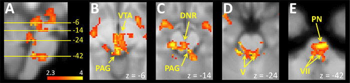
Brainstem regions activated in association with swallowing water when euhydrated
These projects involve standard fMRI as well as perfusion-fMRI. Perfusion-fMRI involves the acquisition
and analysis of arterial spin labelling images to estimate levels of regional cerebral blood flow (rCBF) that can be used to contrast between states (i.e. dehydration, euhydration) separated in time by many minutes or hours. Exercise is used as a strategy to deplete participants of water through increased sweating. The projects address questions about how the brain contributes to the matching of drinking volumes to levels of fluid loss; the influence of hydration status on hedonic aspects of drinking water; and the rapid satiation of thirst in response to drinking despite the relatively slow absorption of the ingested water.
2. Salt Appetite
Not all bodily signals evoke a tangible interoceptive sensation. When depleted of sodium, whether via experimental manipulation or as a manifestation of disease, humans do not experience a craving or hunger for salt. Given the dire implications of hyponatremia, the lack of a human salt hunger does not appear to be compatible with survival. However, the absence of a discernable interoceptive sensation during sodium depletion is possibly redundant because humans have an appetite for salt that leads to levels of consumption far in excess of need. The seeming paradox of excessive salt ingestion combined with the lack of a regulatory hunger for salt has been put thus – “…we will seek salt to please our palate, but not to save our life.” Nevertheless, sodium depletion can occur, despite a strong salt appetite, and there is evidence that the gratifying nature of salt is even more pronounced in the face of a sodium deficit. Consequently, interoceptive processes might impact on sodium levels by modulating the pleasantness of salt and consequently increasing the probability of further ingestion.
These projects will involve measurement of regional brain responses using fMRI, and include acquisition of images optimised for the brainstem. Strategies to deplete participants of sodium include sustained exercise, diet manipulation and administration of diuretics. Ratings of taste intensity (saltiness) and hedonic responses (pleasantness) of salt solutions placed in the mouth will be recorded during sodium replete and depleted states during functional brain image acquisition. These studies will answer fundamental questions about how is sodium depletion is represented in the human brain, and how sodium depletion changes the processing of signals associated with the taste of salt.
3. Temperature Sensations and Thermoregulation
Our sense of temperature has both discriminative and affective components. In respect to discrimination, small changes in skin temperature can be accurately detected and scaled psychophysically, whether they are associated with warming or cooling of the skin. The affective dimension of temperature sensation, or thermal comfort, varies according to the homeostatic implications of the stimulus. For instance, when the core temperature is decreased a cooling skin stimulus will be perceived as unpleasant and a warming stimulus as pleasant. Converse affective responses occur when the core temperature is elevated. The independent nature of discriminative and affective responses to temperature changes makes thermal sensation an excellent model to understand the motivational component of interoception.
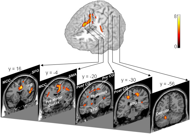
Whole-body heating and cooling is associated with activation in a widely distrusted brain network
These projects use both standard fMRI and perfusion-fMRI to assess regional brain responses to changes in core and skin temperature. A whole-body suit incorporating small tubes connected to a water reservoir is used to change core temperature during scanning. Temperature controlled water is also used to more rapidly apply thermal stimuli to small patches of skin. Experiments using these measures and stimuli will identify brain regions that alternatively code discriminative versus affective components of temperature sensation.
4. Cough and the Urge-to-Cough
Coughing is a vital reflex that protects the airways. Stimuli that evoke cough can also elicit the sensation of an urge-to-cough, which provides an awareness of the airways and can influence discretionary responses such as cough suppression. Understanding how the brain controls cough and represents the urge-to-cough has important implications for pathological conditions where cough is either exaggerated or impaired.
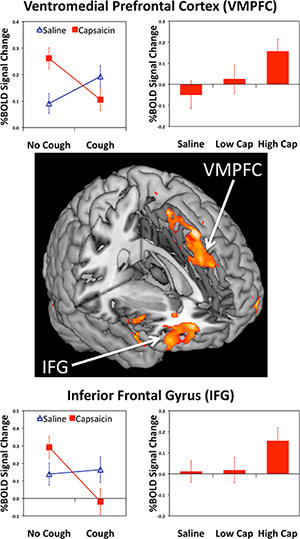
Brain regions implicated in cough suppression include the ventromedial prefrontal cortex and the inferior frontal gyrus.
These projects involve fMRI measures of regional brain responses during the inhalation of tussive (cough promoting) substances, such as nebulised solutions of capsaicin. Doses are titrated according to the sensitivity of the participant and can be adjusted to elicit an urge-to-cough or trigger active coughing. These methods are used to address questions about the central coding of airways inputs, the representation and modulation of urge-to-cough, and the motor control of coughing and cough suppression. Other projects include questions about the neurobiology of placebo effects on the urge-to-cough, and the impact of cough hypersensitivity syndrome on the central processing of airway inputs and coughing control.