Law group
Bioinformatics Research in Artificial Intelligence and Neuroimaging (iBRAIN) research laboratory
Get in touch | Our people | Our work | Our Achievements | Publications
The iBRAIN research group is a large group featuring multiple individual research groups including the Law, Jupp, Harding, Vivash, Sinclair, Spitz, Macefield groups, all with distinct brain disease areas of interest. We use human brain imaging, image processing, and artificial intelligence to develop earlier diagnosis and new treatments for people with brain diseases, and also to find early indications of disease activity (e.g. diagnostic imaging biomarkers). We do this so that patients with brain diseases can be treated earlier and more effectively in clinical practice.
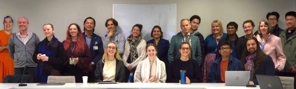
2023 iBRAIN group L-R: Back row: Lucy Vivash, Ian Harding, Sara McPhail, Katherine Kenyon, Meng Law, Mohamad Nazemzadeh, Bianca Jupp, Susmita Saha, Vaughan Macefield, Will Pham, Merran Courtney, Savindu Karunaratne, Imogen Bowden, Lara Fernandez, Joshua Lee, James Liu Front Row: Ella Rowsthorn, Cassandra Marotta, Roxanne Dilcher, Jacob Bunyamin, Ben Sinclair
Get in touch
Whether you want to be involved in our research, you wish to study with us, you want to collaborate with us or donate to our work, we would be delighted to hear from you.
- Email us: meng.Law@monash.edu
Follow us for the latest updates
Our people
Group Leader
Principal Investigators
Our work
We study traumatic brain injury (TBI), epilepsy, neurodegenerative diseases (e.g. Alzheimer’s disease, Ageing, Ataxias, Parkinson’s disease, Multiple Sclerosis, Brain tumours), Mental Health Diseases, Sleep Disturbance by utilising state of the art and ultra-high field magnetic resonance imaging (MRI), positron emission topography (PET), computerised topography (CT), magnetic particle imaging, and photon microscopy approaches of brain structure and pathology. We also house a data warehouse/repository of clinical images for research (Monash iBRAIN – Alfred XNAT imaging databank) and other clinical data types to be used for machine learning, deep learning, artificial intelligence applications in diagnostics and therapeutics. We perform clinical trials on novel therapeutics in neurodegenerative diseases and non-neurological diseases. We have local computing as well as access to MASSIVE, a high performance computing centre at Monash University.
Projects
Our achievements
Grants
- 2022-2024 - Biophysics-informed deep learning framework for magnetic resonance imaging from Australian Research Council (ARC) (Law)
- 2021-2026 - Clinical trial to determine the effects of statins on cognition: STAREE-Mind (Law, Harding)
- 2022-2023 - NIF National Mobile Magnetic Resonance Imaging Network (Law)
- 2021-2025 - Emerging techniques for earlier diagnosis and assessment of severity and progression of artificial stone silicosis (Law)
- 2020-2025 - NHMRC Centre of Research Excellence in Neuroimaging (Law)
Publications
Below are a selection of some of published studies reflecting our high impact work. For a full list of our publications, please visit Pubmed,
- Gradient Patterns of Age-Related Diffusivity Changes in Cerebral White MatterGradient Patterns of Age-Related Diffusivity Changes in Cerebral White Matter. Frontiers in Neurology, 2022. Boban, J. , Thurnher, M. M. , Boban, N. , Law, M. , Jahanshad, N. , Nir, T. M. , Lendak, D. F. & Kozic, D
- Machine learning approaches for imaging-based prognostication of the outcome of surgery for mesial temporal lobe epilepsy. Epilepsia, March 2022. Sinclair, B, Cahill, V, Seah, J, Kitchen, A, Vivash, LE, Chen, Z, Malpas, C, O'Shea M, Desmond P, Hicks R, Morokoff A, King K, Fabinyi G, Kaye A, Kwan P, Berokvic S, Law M, O'Brien T.
- The effect of prolonged spaceflight on cerebrospinal fluid and perivascular spaces of astronauts and cosmonauts. Proceedings of the National Academy of Sciences, April 2022. Barisano, G., Sepehrband, F., Collins, H. R., Jillings, S., Jeurisse B, ... Law M, ... , Floris L Wuyts
- A critical guide to the automated quantification of perivascular spaces in magnetic resonance imaging. December 2022. Pham, W. , Lynch, M. , Spitz, G. , O’Brien, T. , Vivash, L. , Sinclair, B, Law, M.
- Retrospective analysis and prospective validation of an AI-based software for intracranial haemorrhage detection at a high-volume trauma centre. Scientific Reports, 2022 Zia, A., Fletcher, C., Bigwood, S., Ratnakanthan, P., Seah, J., Lee, R., Kavnoudias, H., & Law, M.
- Perivascular spaces as a marker of disease severity and neurodegeneration in patients with behavioral variant frontotemporal dementia. Frontiers in Neuroscience, 2022. Moses, J., Sinclair, B., Schwartz, D. L., Silbert, L. C., O’Brien, T. J., Law, M., & Vivash, L.
- Perivascular spaces as a potential biomarker of Alzheimer’s disease. Frontiers in Neuroscience, 2022 Lynch, M., Pham, W., Sinclair, B., O’Brien, T. J., Law, M., & Vivash, L.
- 18F-FDG-PET hypometabolism as a predictor of favourable outcome in epilepsy surgery: protocol for a systematic review and meta-analysis. BMJ Open, 2022. Courtney, M. R., Antonic-Baker, A., Sinclair, B., Nicolo, J. P., Neal, A., Law, M., Kwan, P., O'Brien, T. J., & Vivash, L.