Using 3D technology to enable the global study of fragile ancient fossils
 This week, Monash researchers published 3D images and digital data of fossils of ancient animals such as monkeys, sabre-tooth cats, elephants and warthogs that lived in South Africa alongside our ancestors. The work – the first of its kind in its size and scope, has been published in the US journal, PLOS ONE.
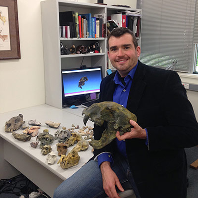
Dr Justin Adams spends months each year in the Cradle of Humankind, 50 kilometres northwest of Johannesburg, South Africa – a region that has produced some of the largest records of early human (or hominin) fossils ever found, some dating back as far as 2.5 million years ago.
Dr Adams, from the Department of Anatomy and Developmental Biology within Monash University's School of Biomedical Sciences, is one of a handful of palaeontologists working on the diverse mammals that lived amongst early humans between 5 million and one million years ago. "Understanding the greater animal community that lived alongside human ancestors helps us understand environments at the time and what ultimately drove the evolution of humans" he says.
As well as producing digital data and models of fossils of ancient animals such as sabre-tooth cats, ancient elephants and warthogs, he has scanned the entire collection of fossil mammals in the vault of South Africa's Ditsong National Museum of Natural History, one of the world's major repositories of ancient fauna fossils, to make them freely available to the world's scientists to compare and study.
"Previously if you wanted to study any of the fossils at the Ditsong, you would have to travel to South Africa and even then, many of the specimens are so fragile that they can't be handled or turned into casts for study," Dr Adams says. The advantage of this 3D digitising research is that the models and CT scans can be downloaded and studied anywhere in the world.
The use of 3D technology and open access to digital data is just starting to revolutionise palaeontology, according to Dr Adams. Several global institutions have started digitising their collections and in June this year the Smithsonian Institute in the US installed its first major 3D printed fossil, a whale.
To digitise a fossil using 3-D technology, a CT or surface scanner captures the shape of the specimen without even touching the original. Surface and laser scanners bounce beams of light off the skull, which reflect back to a sensor on the scanner, dividing the surface up into millions of points that the scanner can reconstruct into a high-resolution surface. Computed tomography (CT) scans, on the other hand, use x-rays, dividing a shape up into cross sections that can be used to see both the outside and inside surfaces of the specimens without damaging it.
When this digital data from surface or CT scanners is combined with 3D printing, new possibilities are opened up for palaeontologists in their research. With a 3D image it is possible to blow up or shrink down the copies it reproduces to highlight areas for study. Digital models can take even a partial fossil, like part of the skull of an ancient hyena or extinct monkey, and used to "create" the rest of the head to allow for measurement and research.
The technology can even take juvenile fossils and create a digital version of what the adult would look like. Take the remains of a famous pre–Homo sapiens hominin discovered in Ethiopia in the 1970s, Australopithecus afarensis, nicknamed Lucy: although dozens of Lucy's bones have been excavated, about 60 percent of the skeleton is missing. To model the complete fossil, researchers have had to reconstruct the missing pieces. If they unearthed the right hand but not the left, for instance, the right limb can be scanned, and the resulting digital template can be flipped into a mirror image, such that a model of the left hand can be printed. The remains of other australopiths in the area also have become digital templates: the vertebrae of a larger individual might be the wrong size for Lucy's frame, but the digital scan can be shrunk to fit, then printed out and fit in place.
For Dr Adams, 3D technology is equally an important tool that will lead to the democratisation of the field. Institutions like the Kenya National Museums have made education-quality scans available to download and, in 2013, the Max Planck Institute started making microCT scans of South African hominin fossils available for researchers.
Dr Adams has also just announced his growing 3D database of scans and CT data of the ancient faunas of the Ditsong Museum in the online journal PLOS ONE, and available for researchers to download through the public data archive MorphoSource.
"With limited funding for field research, this database will provide the first access to these fossils that many scientists have had," Adams says. "Openly sharing this data will encourage further research on these animals and lead to, hopefully, advancements in understanding the context for human evolution."
For those wanting to read the paper in PLOS ONE, click here.