Microscopic beauty of the immune system highlighted
 In the course of an ordinary day, a biomedical researcher might see many images that highlight the hidden beauty of the body under the microscope, such as when looking at cells, the immune system, or the nervous system. Now, three of our researchers have shared the beautiful secrets of the immune system in an image competition for the Melbourne Day of Immunology - Snapshots of the Immune System Scientific Photography Exhibition 2016.
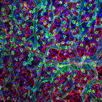
Dr Samantha Dando took out the top spot in the competition with her image (main image) of resident immune cells within the central nervous system – the microglia. Dr Cecilia Naranjo Golborne and Professor Paul McMenamin also had their image (below) of the surface of a brain published in Cosmos Magazine as part of a feature on Melbourne Day of Immunology.
More information on both images is given by the researchers below.
Dr Dando's image of the neural retina contains a rich population of microglia, which are the resident leukocytes within the central nervous system (CNS). In steady-state conditions, motile microglial processes constantly survey the tissue for pathogens and debris, and these cells play an important role in maintaining the immune privileged microenvironment of the CNS. In the CX3Cr1gfp/gfp mouse retina, microglia within the outer plexiform layer are visualised by their expression of green fluorescent protein. Blood vessels (red) were stained with isolectin-B4 and nuclei (blue) were stained with Hoechst. This image was captured using a 40x objective and a Leica SP5 confocal microscope at Monash Micro Imaging, Monash University.
 Dr Cecilia Naranjo Golborne and Professor Paul McMenamin's image (left) of a confocal microscopy study of the surface of the Gray short-tailed opossum (Monodelphis domesticus) brain stained with lectins and antibodies shows a network of blood vessels and myeloid cells within the pia mater. The pia mater is the inner-most layer of the meninges, which protects the brain. The macrophages in the pia mater and its blood vessels are labelled with isolectin-B4 (green). Underlying the pia mater, anti-glial fibrillary acidic protein staining reveals a dense network of astrocytes (red) in the superficial cerebral cortex of the brain. Microglia, the resident immune cells of the central nervous system parenchyma, are also present within the cerebral cortex and express ionised calcium-binding adapter molecule-1 (magenta). Nuclei (blue) are stained with Hoechst. This image was captured using a 20x objective and a Leica SP5 confocal microscope at Monash Micro Imaging, Monash University.