
New 3D model reveals hidden driver of lung scarring, paving path to new treatments
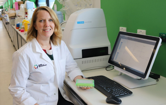
Associate Professor Katrina Binger.
Researchers have uncovered a previously unknown mechanism that may contribute to idiopathic pulmonary fibrosis (IPF), a progressive and often fatal lung disease with limited treatment options.
Published in Science Advances, the research reveals how a protein found in damaged tissue - vitronectin - can reprogram immune cells leading to the formation of scar tissue in the lungs that causes pulmonary fibrosis.
Fibrosis, the excessive build-up of scar tissue, contributes to diseases affecting the lungs, heart, kidneys and other organs. In pulmonary fibrosis, this scarring gradually reduces the lungs’ ability to deliver oxygen to the body. Although immune cells known as macrophages are known to play a central role in the disease, researchers have struggled to understand exactly what transforms them into drivers of tissue damage.
Led by Associate Professor Katrina Binger at Monash University’s Biomedicine Discovery Institute, with her collaborators at La Trobe Institute for Molecular Science, the team tackled this challenge by moving beyond conventional laboratory methods, which typically study cells on flat plastic surfaces. Instead, the team developed a three-dimensional model that better reflects the environment cells experience inside the body, embedding macrophages within a collagen-rich matrix containing vitronectin.
Using this more physiologically relevant system, the researchers discovered that vitronectin fundamentally alters macrophage metabolism, increasing expression of the enzyme CD38 and driving the cells towards a pro-fibrotic state. These changes were not apparent in traditional two-dimensional cultures, highlighting how important tissue architecture can be in shaping immune cell behaviour.
“We normally think of vitronectin as a structural protein that maintains the integrity of organs like the lungs. But we found it also can also act as a signal,” Associate Professor Binger said.
“We found that in these 3D environments, vitronectin changes how macrophages produce energy, and this drives them to have a heightened fibrotic state.
“This is a completely new mechanism to understand how fibrosis happens that was only possible by studying these cells in more natural, 3D environments,” she said.
The findings were validated in both human and animal studies. Lung tissue from patients with IPF contained elevated levels of vitronectin and increased numbers of macrophages carrying the same molecular signature identified in the laboratory model. In addition, mice lacking vitronectin were protected from experimental lung fibrosis, showing reduced scarring, less inflammation and improved lung function.
Associate Professor Binger said the study demonstrates that the physical and molecular environment surrounding immune cells can be just as important as the cells themselves in driving disease. By identifying vitronectin and macrophage metabolism as key contributors to fibrosis, the research provides promising new targets for future therapies and underscores the value of advanced 3D models for uncovering disease mechanisms that would otherwise remain hidden.
A quick visual walkthrough of our paper’s core discoveries. Watch how a macrophage cell’s internal "battery" becomes fully charged upon interacting with vitronectin and collagen in a 3D matrix, triggering the progressive, dense scarring cascade that drives lung fibrosis. Concepts, storyboarding, and video editing by Katrina Binger. Visual assets and micro-animations generated using Adobe Firefly.
Read the full paper published in Science Advances, titled Vitronectin metabolically programs pro-fibrotic macrophages in 3D cultures and idiopathic pulmonary fibrosis
MEDIA ENQUIRIES
For more Monash media stories, visit our news and events siteMonash Media
E: media@monash.edu
T: +61 (0) 3 9903 4840
About the Monash Biomedicine Discovery Institute at Monash University
Committed to making discoveries that will relieve the future burden of disease, Monash Biomedicine Discovery Institute at Monash University brings together more than 120 internationally renowned research teams. Spanning seven discovery programs across Cancer, Cardiovascular Disease, Development and Stem Cells, Infection, Immunity, Metabolism, Diabetes and Obesity, and Neuroscience, Monash BDI is one of the largest biomedical research institutes in Australia. Our researchers are supported by world-class technology and infrastructure, and partner with industry, clinicians and researchers internationally to enhance lives through discovery.