News & events
 | Alex Fornito awarded ARC Laureate Fellowship30 August 2022 We are thrilled to announce that Professor Alex Fornito has been awarded a prestigious ARC Laureate Fellowship for 5 years titled "Next-generation maps and models of the human brain". The scheme supports exceptional, world-class researchers to undertake groundbreaking and transformational research and to foster an excellent research training environment and exemplary mentorship to nurture early-career researchers. This year, in total 16 fellowships were awarded across the country, making them extremely competitive. |
 | Focused ultrasound for treating brain tumours13 April 2022 Finding an effective treatment for brain tumours has challenged researchers for decades, partly due to the impenetrability of the Blood Brain Barrier (BBB). Focused Ultrasound (FUS) enhances drug delivery and accumulation to infiltrating tumour regions by inducing targeted, temporary disruption of the BBB. Monash Biomedical Imaging's Dr Ekatarina Salimova is using this innovative technique to find new treatment targets in preclinical models of glioblastoma. Dr Salimova and colleagues have recently published a comprehensive review paper on the current translational status of the preclinical FUS research in the Journal of Controlled Release. |

| $71M Australian Precision Medicine Enterprise project4 April 2022 Monash Biomedical Imaging is part of the new $71.2 million Australian Precision Medicine Enterprise (APME) facility, which brings together industry partners Global Medical Solutions Australia (GMSA) and Telix Pharmaceuticals with Monash University. The APME will deliver large-scale development and manufacturing of precision medicines and theranostic radiopharmaceuticals for industry and research. The project has received $23 million from the Australian Federal Government. A key feature of the APME project is a high-energy cyclotron with multiple production clean rooms, which will be located on the Monash Biomedical Imaging site in Clayton. This strategic co-location will facilitate radiochemistry, PET and SPECT research and clinical use of theranostic (therapeutic and diagnostic) radioisotopes produced on-site. Read the Monash University announcement. Visit the APME Website and LinkedIn. |

| Triceratops comes to life with CT imaging16 March 2022 Computer Tomography (CT) scans of a 67-million-year-old Triceratops acquired at Monash Biomedical Imaging have been used to create an immersive digital experience as part of the dinosaur’s exhibition at the Melbourne Museum. Data from the scans produced by MBI’s large bore Siemens CT scanner will also be digitally archived and maintained by the Museum. |

| CT plus microbeam radiation therapy to treat brain cancer27 October 2021 Combining high-precision CT imaging with microbeam radiation therapy could lead to improvements in personalised treatments for brain cancer and reduce damage to healthy tissue that can be caused by current radiation treatments. New research published in MDPI Applied Science has demonstrated a novel method of targeted microbeam radiation therapy using CT imaging and the Australian Synchrotron's medical beamline. The study used both of Monash Biomedical Imaging's large and small bore CT scanners for optimum CT imaging to identify the tumour site and allow 3-dimensional targeting in rats. Then patient-specific CT-guided microbeam radiation therapy with the medical beamline was used to treat the tumour. The radiation was delivered well within the margins of acceptable treatment area and did not destroy healthy tissue. |

| Prestigious PhD excellence award for MBI student9 August 2021 A prestigious award from the Indian Institute of Technology Bombay has been awarded to Dr Viswanath Pamulakanty Sudarshan, who recently completed his PhD research at Monash Biomedical Imaging. He received the ‘Naik and Rastogi Award for Excellence in PhD Research’ for 2019-2021, which was presented by Professor Geoffrey Hinton from the University of Toronto. Read more. |

| Attentional lapses are linked to local sleep-like activity in the awake brain5 July 2021 Dr Thomas Andrillon and Professor Nao Tsuchiya from MBI's Linked Lab, Monash Neuroscience of Consciousness, have discovered that the presence of slow waves in the awake brain is linked to lapses in attention such as mind-wandering and mind-blanking. Read more. |

| User Access Scheme grants awarded22 June 2021 This year we introduced the Monash Biomedical Imaging (MBI) and Alfred Research Alliance–Monash Biomedical Imaging (ARA-MBI) User Access Scheme to promote, facilitate and support high quality research involving the use of the imaging services and expertise at MBI in Clayton and ARA-MBI in Prahran. The scheme was open to all Monash University and Baker IDI researchers. Find out this year's User Access Scheme grant recipients. |
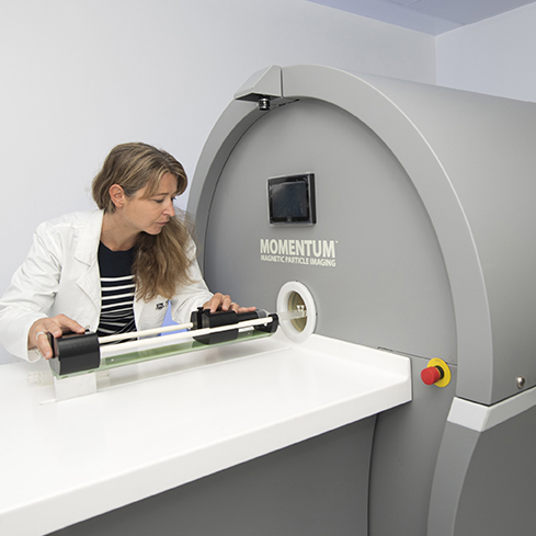
| A world first in imaging technology15 June 2021Monash University has launched a world-first technology that can detect magnetic nanoparticles anywhere in the body, enabling enhanced medical applications such as tracking of beneficial CAR-T cells during cancer therapy. The new technology combines Magnetic Particle Imaging (MPI) with Computed Tomography (CT) and Hyperthermia capabilities for preclinical, in vivo research. Read more. |

| Virtual Launch of Magnetic Particle Imaging at Monash UniversityDATE: Tuesday 15 June 2021 The Magnetic Particle Imaging capability at the Alfred Research Alliance - Monash Biomedical Imaging site is the world’s first MPI system with Computed Tomography (CT) and Hyperthermia capabilities. It provides unique capabilities that open the door to cutting-edge opportunities for interdisciplinary projects in medical research, chemistry and biotechnology. Further details and to register for this virtual event: https://mpi.eventbrite.com.au |

| Prestigious global scholar award26 May 2021Associate Professor Adeel Razi has been selected as a prestigious CIFAR Azrieli Global Scholar. A/Prof Razi leads the Computational and Systems Neuroscience Linked Lab at Monash Biomedical Imaging. A/Prof Razi will use MBI’s brain imaging facilities to conduct his CIFAR research, which aims to uncover the neural mechanisms underlying altered states of consciousness and how these insights may help build general-purpose artificial intelligence. He will be part of the Brain, Mind and Consciousness program while his research will also include close interactions with the Learning in Machines and Brains program at CIFAR. The CIFAR program is cross-disciplinary and will see A/Prof Razi work with other early career researchers from the social and natural sciences. The CIFAR Azrieli Global Scholar program is a highly selective program with recipients selected based on their research excellence and potential to be leaders of tomorrow. |

| CT helps researchers learn how seals evolved their flippers to optimise swimming7 May 2021MBI's large bore CT scanner was used in a cross-discipline study to discover why some seals and sea lions swim differently to others. Seal and sea lion flipper specimens were scanned with MBI's Siemens Somatom GoUp CT scanner. The images were refined by MBI's Preclinical Support team so that the researchers could use these images to create cutting-edge computer simulations. By combining the 3D simulations alongside footage of live seals, the researchers answered this evolutionary mystery. The Monash University-led study included researchers from the School of Biomedical Sciences and the Department of Mechanical and Aerospace Engineering, as well as researchers from University of Otago and Macquarie University. Read more. |
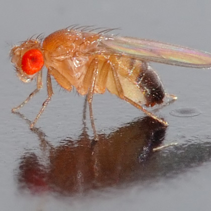
| Testing a theory of consciousness in flies15 March 2021 Tests in fruit flies show that “informational structures” of the brain could be used to measure levels of consciousness. Read more about this new research from Monash Biomedical Imaging Linked Lab, Monash Neuroscience of Consciousness. |

| New MRI technique for neurodegenerative disease18 February 2021 In amyotrophic lateral sclerosis (ALS), also known as motor neuron disease, iron accumulation in the motor cortex is a pathological marker of the disease. Monash Biomedical Imaging's Imaging Analysis team has shown that quantitative iron assessment in the brain using recent advances in MRI, including quantitative susceptibility mapping (QSM), holds great potential to be a sensitive diagnostic and prognostic marker in ALS. Read the Journal of Magnetic Resonance Imaging research review article. |

| Measuring changes in attention, not perception21 January 2021 Neuroscience has long grappled with the relationship between attention and conscious awareness, or perception. Research from MBI Linked Lab investigators suggests that the two processes are separate and use different pathways in the brain. This is illustrated by studies showing that we can be conscious of things we’re not paying attention to, and not conscious of things we are paying attention to. Read more. |
| Neuroscience in a Flash winner14 December 2020 PhD student Winnie Orchard from the Cognitive Neuroimaging team won the People’s Choice Award in the ‘Neuroscience in a Flash’ competition, held by the Faculty of Medicine, Nursing and Health Sciences at Monash University. For the competition, graduate research students were required to creatively present their research thesis in three minutes, using only one slide. |
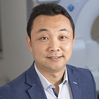
| Zhaolin Chen awarded ARC Discovery Project17 November 2020 Head of MBI’s Imaging Analysis team, Dr Zhaolin Chen, has been awarded a 2021 ARC Discovery Project grant for his research into biophysics informed artificial intelligence in MRI image acquisition and reconstruction. Learn more about Zhaolin Chen's research. |
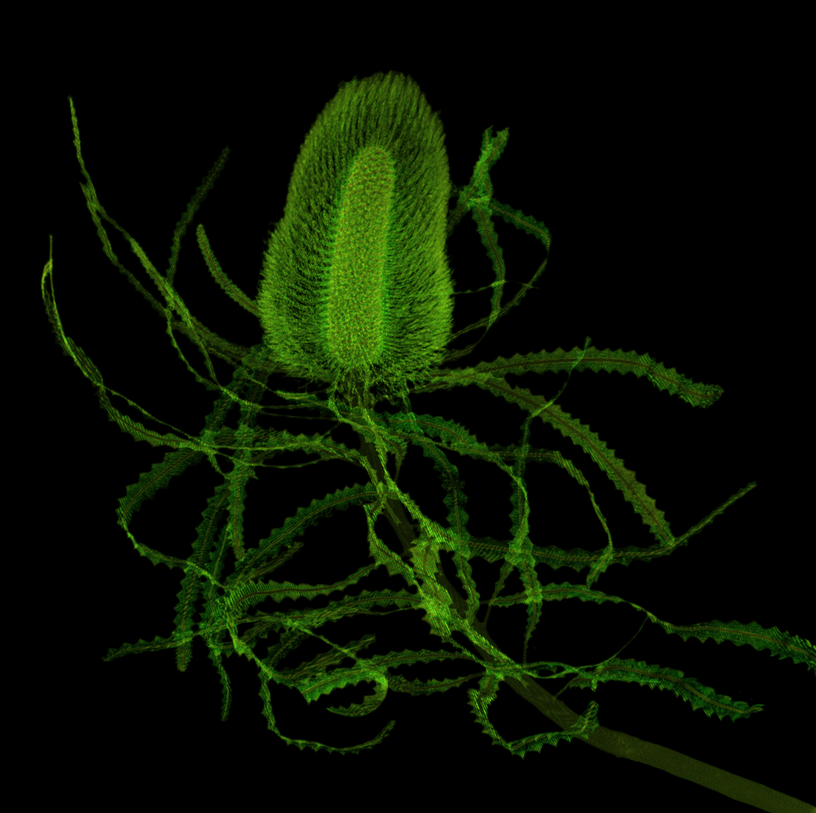
| Exploring bio-aesthetics using CT imaging9 November 2020 Using CT imaging capabilities at Monash Biomedical Imaging, award-winning artist Valerie Sparks is exploring ways to collect, manipulate and present 3D datasets. Her art project in progress, named ‘Bio-aesthetic’, focuses on the rarely visualised interior of flora. Read more from the National Imaging Facility on this work. |
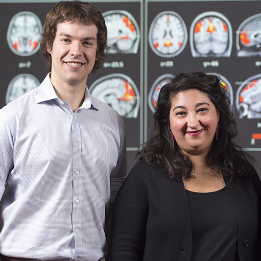
| A new open dataset for studying the brain3 November 2020A new publicly accessible Monash rsPET-MR dataset will help researchers to understand network dynamics in the brain. Developed by Monash Biomedical Imaging researchers, Dr Sharna Jamadar and Dr Phillip Ward, researchers in the brain imaging community can use this unique dataset to understand the relationship between oxygen and glucose use during dynamic brain function. They can also use it to develop new methods and scientific discoveries. Read more. |

| Many women feel ‘phantom kicks’ after pregnancy15 October 2020 The prevalence of ‘phantom kicks’ after the end of pregnancy has implications for fetal health monitoring and women’s mental health. This research, published in the Journal of Women's Health, was led by Dr Sharna Jamadar from MBI's Cognitive Neuroimaging team. Read more. |
| Haemoglobin levels affect the results of brain connectivity studies11 September 2020 Natural variations in haemoglobin levels should be considered when using functional MRI to study brain connectivity. This discovery was made by researchers from MBI's Cognitive Neuroimaging team and also the Neural Systems and Behaviour Lab. Read more. |
| Parenthood permanently changes the brain27 August 2020 Research from Monash Biomedical Imaging's Cognitive Neuroimaging team has shown the experience of raising children is linked to life-long changes to the brain’s structure in mothers and fathers. Read more. |
| MBI researcher awarded ISMRM Junior Fellow10 August 2020 Monash Biomedical Imaging (MBI) researcher, Dr Phil Ward, has been selected as a Junior Fellow of the International Society for Magnetic Resonance in Medicine (ISMRM), the preeminent association for researchers who specialise in magnetic resonance imaging (MRI). Read more. |
|
| Outstanding results for two of MBI's young researchers30 July 2020 Dr Phillip Ward and PhD student Viswanath Pamulakanty Sudarshan (pictured) were recently recognised for their outstanding research at the International Society for Magnetic Resonance in Medicine (ISMRM) annual meeting. The meeting had over 5000 lectures and many more posters. One of Dr Ward’s abstracts placed in the top 5% of those submitted by early career researchers, and another in the top 15%, while one of Viswanath’s abstracts ranked in the top 15% category. |

| MRI method improves fPET analysis13 July 2020 New research from MBI's Imaging Analysis team shows how a magnetic resonance imaging method improves functional PET (fPET) analysis. The magnetic resonance-guided fPET framework reduces partial volume errors, enhances the sensitivity of identifying brain activation, and improves the anatomical accuracy for mapping changes of brain metabolism in response to a visual stimulation task. The results demonstrate that the framework extends the use of fPET to investigate the dynamics of brain metabolic responses for faster presentation of brain activation tasks, and for applications in low dose PET imaging. |

| New brain imaging research could help improve motor rehabilitation29 June 2020 Our central nervous system is constantly faced with the challenge of adapting to rapid fluctuations in our environment. Dr Michelle Marneweck, who is based at Monash Biomedical Imaging, has discovered how the brain learns to anticipate and correct for such fluctuations, specifically when it comes to dexterously lifting objects. As part of her multimodal research approach, Dr Marneweck and her colleague Dr Scott Grafton from the University of California, Santa Barbara used rapid multiband brain imaging, in conjunction with kinematics and Bayesian pattern component modelling. This discovery contributes to our understanding of how the brain enables dexterous action in the face of rapidly fluctuating dynamics, which has significant implications for the advancement of human brain-machine interfaces and motor rehabilitation. Her research was recently published in Nature Scientific Reports. |
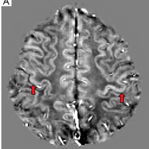
| Neurodegenerative disease research published9 June 2020 New research from MBI’s Imaging Analysis team may help improve our understanding of Amyotrophic Lateral Sclerosis (ALS), a fatal neurodegenerative disease. This study demonstrates the efficacy of Quantitative Susceptibility Mapping in the detection of susceptibility changes, possibly due to iron dysregulation in the motor cortex in ALS. These findings may lead to the development of a sensitive neuroimaging biomarker that can provide meaningful insight of pathophysiologic changes in ALS subtypes. |
| MBI – Synchrotron research in Nature Scientific Reports4 June 2020 Researchers from the University of Wollongong have published research in Nature Scientific Reports on personalised synchrotron microbeam radiation therapy (MRT) for brain cancer treatment. As part of this research, rat models underwent CT imaging with Monash Biomedical Imaging's Siemens Inveon PET/CT scanner to visualise tumour against normal brain tissue before MRT. |
|
| NHMRC Investigator Grants for MBI collaborators20 May 2020 Prestigious NHMRC Investigator Grants have been awarded to a number of researchers connected with MBI. Dr Adeel Razi (pictured), from our Linked Lab Computational Systems and Neuroscience received a grant to develop mathematical models for tracking early stage dementia using non-invasive brain imaging. Professor Alex Fornito from the Brain, Mind and Society Research Hub, co-located at MBI, has received a grant for his project, ‘A network approach to mapping and modifying brain changes in psychosis’. Dr Rico Lee from BrainPark, also co-located at MBI, will use his grant to research a purpose-built digital assessment tool to determine the mechanisms driving addictive behaviours and its utility to improve treatment engagement and outcomes. |
|
| Deep learning improves imaging for prostate cancer14 May 2020 Monash Biomedical Imaging's Imaging Analysis team has published research using novel deep learning methods to improve MR-PET imaging of prostate cancer. Compared with the traditional MR based PET attenuation correction methods, the team’s new deep learning techniques improved the accuracy of attenuation correction for MR-PET images and led to more accurate PET quantification in a cohort of 28 prostate cancer patients. Read the team’s paper, published in the European Journal of Nuclear Medicine and Molecular Imaging. |
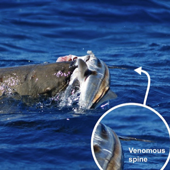
| Deadly seafood: fish use sharp barbs and spines to fight off hungry seals4 May 2020 Using Monash Biomedical Imaging's large bore CT scanner, researchers from Monash University have revealed the steep price some marine mammals are willing to pay for food, after a stranded fur seal was discovered with more than a dozen facial wounds inflicted by its seafood prey. |

| New method produces faster and higher quality human imaging2 April 2020 Researchers from Monash Biomedical Imaging have developed a way to combine magnetic resonance and positron emission tomography (MR-PET) data through a joint image reconstruction system that also improves the quality and speed of images generated by simultaneous MR-PET scanners. |

| New methods developed for MR imaging of lung13 February 2020 Magnetic resonance imaging of lung tissue has previously been difficult to achieve but the recent development of using hyperpolarised xenon as a contrast agent has revolutionised the field of functional lung imaging. Now MBI researchers have developed a machine that can reliably produce hyperpolarised xenon for MR lung imaging. |
|
| Monash researchers in consortium granted $1million for lung disease research10 February 2020 Monash Biomedical Imaging will provide advanced MR-PET imaging expertise for a project that has the potential to improve treatments for the lung disease idiopathic pulmonary fibrosis (IPF). |

| MBI team leader attends prestigious PM awards17 October 2019 Leader of MBI's Cognitive Neuroimaging Team, Dr Sharna Jamadar, attend the Prime Minister's Prizes for Science awards ceremony in Canberra last night. These prizes are Australia’s most prestigious science awards and last night's event celebrated 20 years of the PM’s Prizes. |

| An image of health: detecting brain damage in high risk babies14 October 2019 The impact of a serious pregnancy condition that affects millions of unborn babies globally could be minimised by improved imaging techniques. Hudson Institute of Medical Research and Monash Biomedical Imaging scientists used advanced imaging techniques to analyse magnetic resonance imaging (MRI) scans to detect subtle brain injury associated with foetal growth restriction (FGR), a condition that leads to babies not growing optimally in the womb. |

| Travel Award assists imaging and deep learning knowledge12 September 2019 Rebekah Chiu, a student with the Imaging Analysis Team, has been awarded a Medical Image and Computing and Computer Assisted Intervention (MICCAI) Travel Award to attend the 2019 MICCAI Conference in Shenzhen, China. As part of her visit she will access additional tutorials and workshops, and experience many opportunities to network and learn from experts in imaging and deep learning. |
|
| Early career researcher wins award for MRI research5 September 2019 Dr Phil Ward from the Cognitive Neuroimaging Team has been awarded the Jenny Redman Early Career Researcher Publication Prize for Psychological Sciences, Monash University, for his paper 'Combining images and anatomical knowledge to improve automated vein segmentation in MRI', which was published in NeuroImage. |
|
| MBI researchers receive prestigious investigative grants3 September 2019 Congratulations to Dr Sharna Jamadar and Dr Phil Ward from MBI's Cognitive Neuroimaging Team on receiving prestigious NHMRC Investigator Grants. This funding secures their research for the next five years. Their work on healthy ageing using MBI's simultaneous MR-PET scanner is world-leading. |
| Advanced MRI analysis detects brain injury in foetal growth27 August 2019 New research published in NeuroImage shows the effects of foetal growth restriction on brain development. The MRI scanning and analysis for this research was conducted at Monash Biomedical Imaging using the 3T MRI Skyra. |
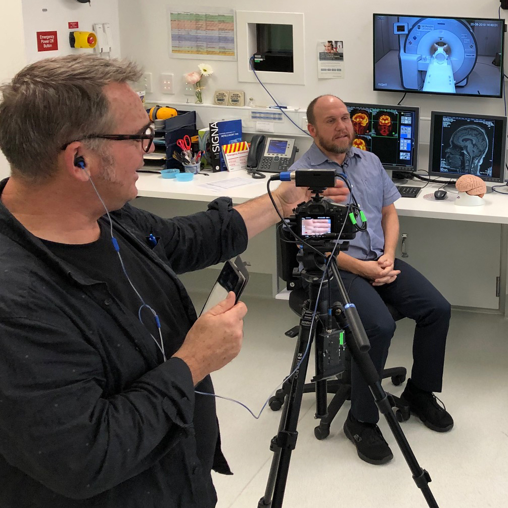
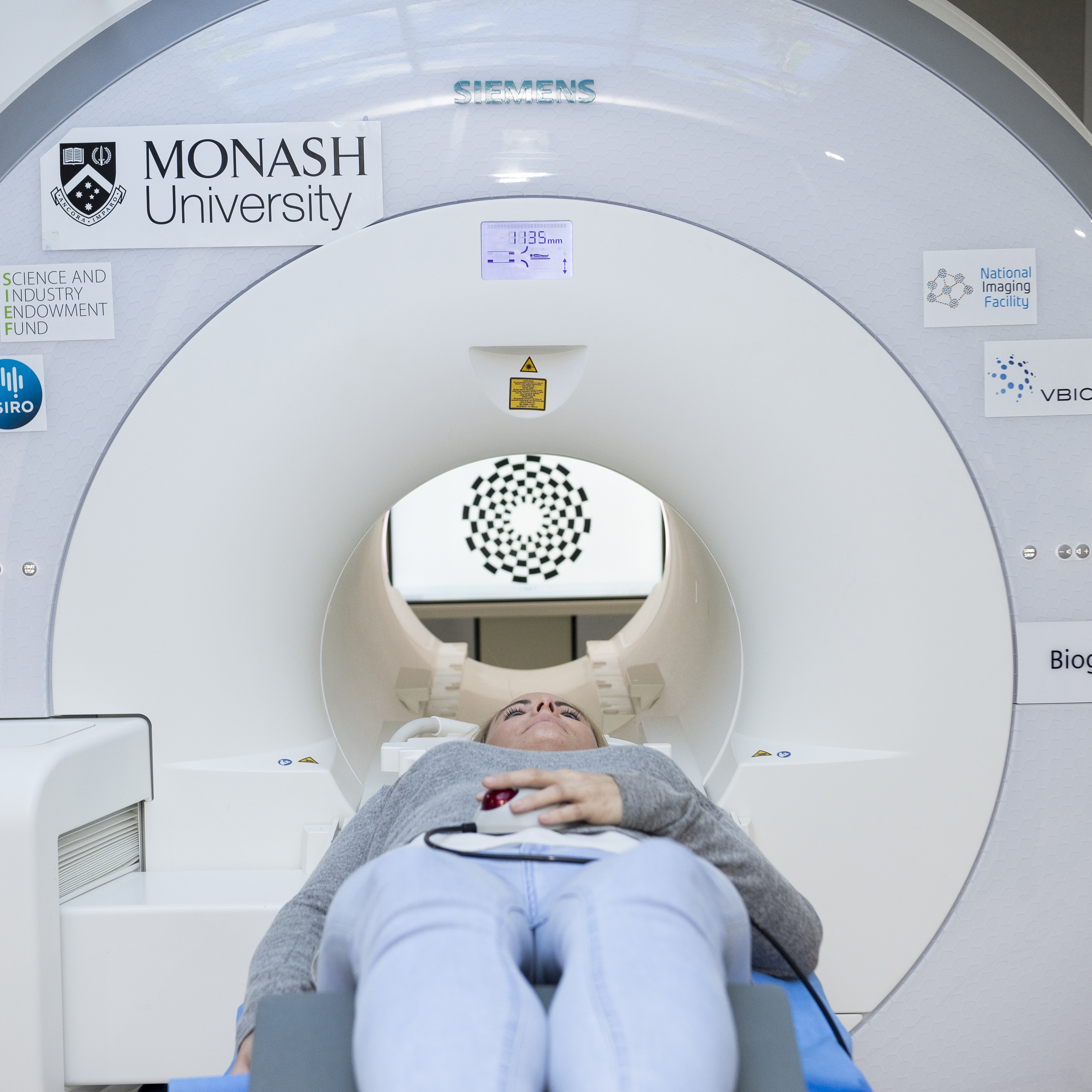
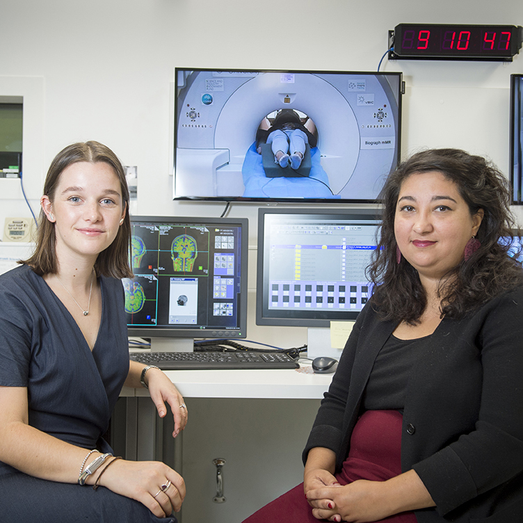
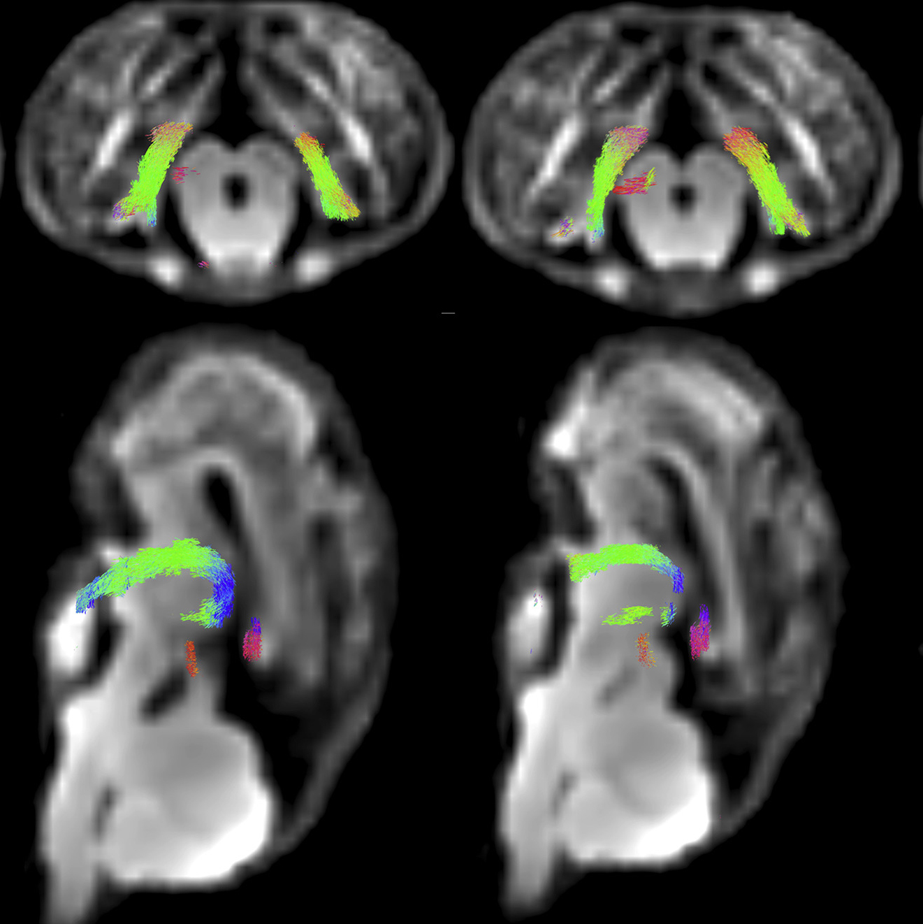
| MR-PET procedure filmed for visual journal20 August 2019 Dr Sharna Jamadar and her Monash Biomedical Imaging colleagues spent a whole day filming one of their MR-PET imaging procedures for the Journal of Visualised Experiments. The team were filmed conducting the procedure, 'Radiotracer Administration for High Temporal Resolution Positron Emission Tomography of the Human Brain: Application to FDG-fPET'. The paper is currently available online with the video soon to follow. |
|
| Precision medicine to be advanced by international collaboration15 August 2019 Individualised precision medicine to treat a range of human diseases will be the focus of the newly formed Monash University Network of Excellence for Molecular Imaging and Precision Radiopharmaceuticals. |
| How Much Water Do You Really Need to Drink?17 July 2019 Myth busted! You DON'T need to drink 8 glasses of water a day, according to Monash Biomedical Imaging's Associate Professor Michael Farrell. “Drink when you want to, and chances are this behaviour will keep your fluid balance on an even keel.” Read why Associate Professor Farrell gives this advice in Elemental. |

| Towards the physical basis on consciousness2 June 2019 Questions about consciousness have puzzled humanity for centuries. In a recent Convergence Science Network event held at Monash Biomedical Imaging, Associate Professor Nao Tsuchiya from the Monash Neuroscience of Consciousness group discussed the latest neuroscience research on consciousness. These scientific discoveries may help answer the philosophical questions about consciousness. View the event. |
| Renowned cardiologist to lead Victorian Heart Hospital and expand research28 May 2019 World renowned cardiologist and researcher Stephen Nicholls will lead the new Victorian Heart Hospital, and expand his research at Monash University. Monash Biomedical Imaging (MBI) will be a key partner in the Hospital's research and innovation, with its researchers accessing MBI's latest preclinical imaging facilities. |
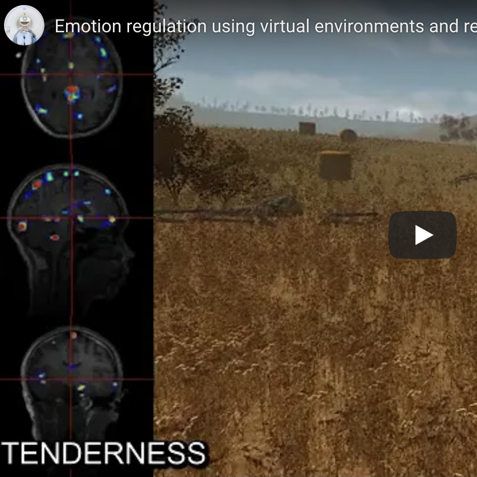
| Viewing and changing emotions in real-time with new brain imaging tool2 April 2019 A world-first technology that combines virtual reality, a brain computer interface and magnetic resonance imaging is providing new insights into brain activity during complex emotional states. |
|
| Trippy tech explores brain power under LSD1 April 2019 Monash University researchers are part of a global team that has developed and applied world-first non-invasive computer technology to explore how the brain processes information when under the influence of hallucinogens, like LSD. |

| Breakthrough discovery further suggests attention and consciousness are separate27 February 2019 When we focus our attention on one thing, our brains rapidly change attentional focus. Even though we may feel like we’re paying full attention to something, these rhythmic brain mechanisms suggest our brain itself never stops searching for other things to attend to. |

| EEG research shows our attention can jump between visible and invisible objects13 December 2018 Monash University researchers have used MBI’s EEG facilities to investigate how attention is focused and diverted, and how attention interacts with what we consciously perceive. |

| MBI scientist selected for Superstars of STEM11 December 2018 Dr Sharna Jamadar from Monash Biomedical Imaging's Cognitive Neuroimaging Team has been selected to join the prestigious 2019/20 Superstars of STEM program. Entry into the program is highly competitive and Dr Jamadar will spend the next two years undertaking activities that aim to remove society’s gender assumptions about scientists and increase the public visibility of women in STEM. |

| MRI helps diagnose Leadbeater's Possum illness4 December 2018 A Leadbeater's Possum from Healesville Zoo was behaving outside his normal routine, so a team of veterinary specialists and imaging experts from Monash Biomedical Imaging worked together to find out what was causing this possum to be unwell. Using MBI's small animal MRI scanner, the team discovered lesions on the possum's brain and he was diagnosed with a neurological condition. Treatment for 'Phoenix' the possum commenced straight away and he is now responding well and being closely monitored. |

| ARC grant for Australia's first Magnetic Particle Imaging facility30 November 2018 MBI Director, Professor Gary Egan, will be the lead investigator with Australia's first Magnetic Particle Imaging (MPI) facility. Professor Terry O'Brien, Head of the Department of Neuroscience and Associate Professor Christoph Hagemeyer, head of the NanoBiotechnology Laboratory at the Australian Centre for Blood Diseases at the Central Clinical School will be Chief Investigators. The newly funded MPI machine, along with the recently installed 9.4T MRI, PET-CT and high resolution structure CT-FLECT machines, will make the Alfred Research Alliance (ARA) Preclinical Imaging Facility, led by Dr David Wright, the most advanced in the country, and one of the leading such facilities in the world. |

| Ultrasound imaging for aortic aneurysm preclinical drug trial6 November 2018 Aortic aneurysms are a degenerative disease that generally occur in the abdominal region and affect people over the age of 65 years. They are a silent killer with a mortality rate over 90 percent. Researchers from Monash Biomedicine Discovery Institute’s (BDI) Cardiovascular Disease group are using Monash Biomedical Imaging’s Vevo2100 Ultrasound in their preclinical trials of novel drugs to stabilise established abdominal aortic aneurysms. |

| Computational neuroscience takes brain research in new direction23 October 2018 Improved treatments for debilitating brain disorders may only be possible through interdisciplinary research efforts that work towards the common goal of improving our understanding of the brain. A new research group located at Monash Biomedical Imaging (MBI) is using computational neuroscience to investigate neurodegenerative and neurodevelopmental disorders. Computational neuroscience uses mathematical models and theoretical analysis to find new ways to understand the brain’s working mechanisms. |

| Tackling compulsive behaviours with a playground for the brain11 September 2018 |

| The obesity epidemic: how the brain responds to food choices27 March 2018 View the YouTube video or read the full article on the Monash Lens. |

| MBI imaging expert a key speaker at international conference22 March 2018One of Monash Biomedical Imaging’s research fellows will deliver three oral presentations at the world’s biggest conference for imaging scientists later this year. Dr Kamlesh Pawar will present at the International Society of Magnetic Resonance in Medicine meeting in Paris, 16-21 June. His presentations will include his research on novel pulse sequence development and deep learning based image reconstruction methods, which aim to improve the image quality and reduce the scan time of MR imaging. |

| Creating next-gen neuroimaging technologies6 March 2018Monash Biomedical Imaging's Prof Gary Egan, Dr Sharna Jamadar and Dr Zhaolin Chen have received an ARC Linkage Grant to develop new techniques to improve the data produced by MR-PET scanners. Their work to better fuse the MR and PET data will enable researchers to discover new aspects of the brain that are not possible using each technology alone. |

| How your brain stops you from drinking too much28 January 2018 Monash Biomedical Imaging's Prof Gary Egan and A/Prof Michael Farrell, along with researchers from the Florey Institute of Neuroscience and Mental Health, have used magnetic resonance imaging to discover the area of the brain that regulates our fluid intake and tells us when to stop drinking by predicting when we have consumed enough to rehydrate. |

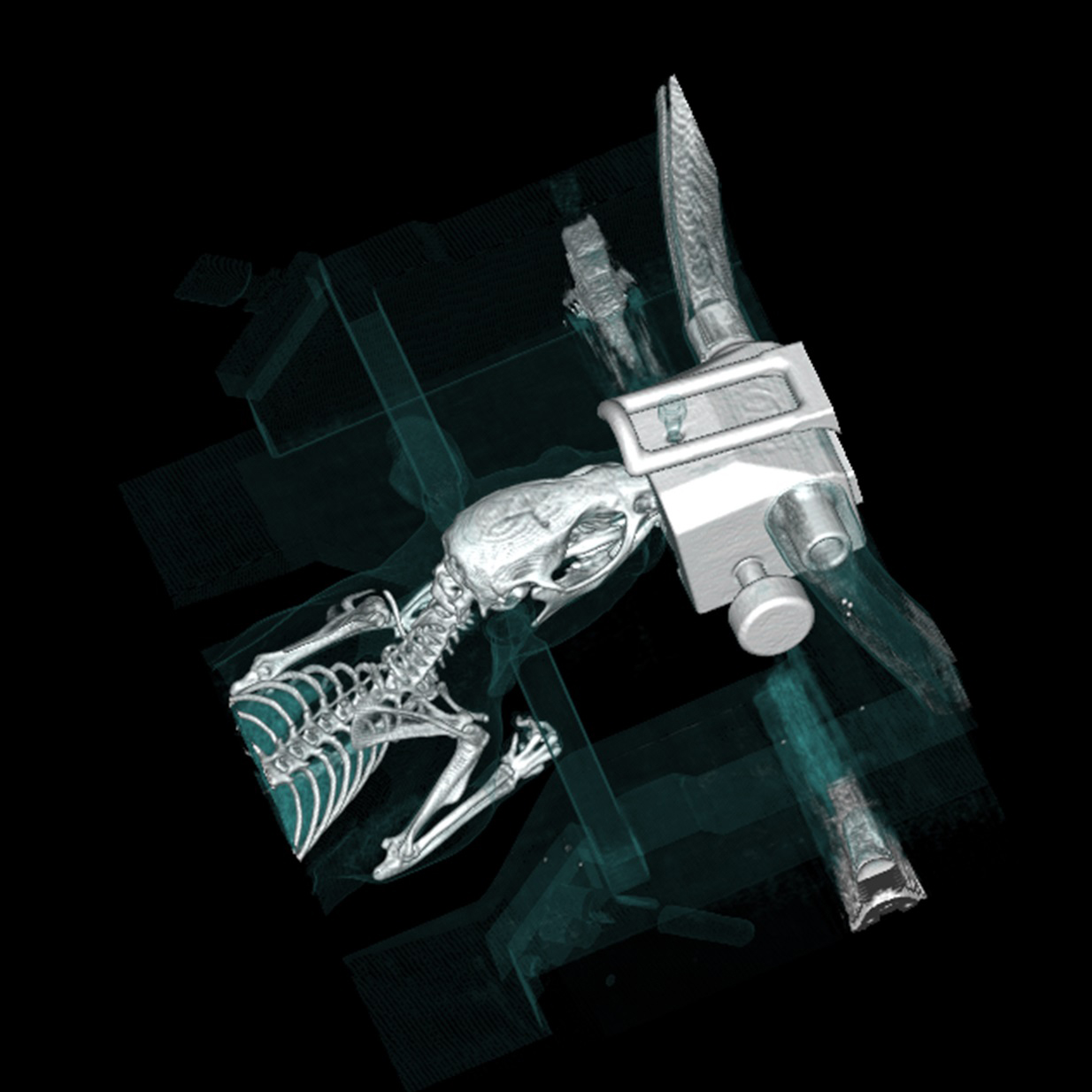
| New CT scanner brings extinct animals to life19 December 2017 Monash Biomedical Imaging’s new CT technology is helping researchers to digitally archive extinct Australian animals, while other investigators are using the scanner’s high-resolution power to develop realistic 3D surgical training simulators. |

| Australia’s first dedicated heart hospital17 December 2017 The Victorian Heart Hospital is a partnership between the Victorian State Government, Monash University and Monash Health. It is a Victorian flagship health project, which will deliver world-leading cardiac services and boost the Victorian economy through innovation, education and research. Monash Biomedical Imaging will be a key partner in the Hospital's research and innovation. |

| Clinical imaging data access and storage improved12 December 2017 Magnetic resonance imaging (MRI) scanners are expensive equipment capable of producing large amounts of valuable research data. For investigators to maximise their research outcomes, the data they obtain from MRIs must be stored securely, its quality verified, and it should be accessible to the wider research community. |

| Victoria Fellowship awarded to MBI researcher31 October 2017 Monash Biomedical Imaging's Dr Phil Ward was one of 12 early career researchers to receive a Victoria Fellowship this year. He will use the award to study new magnetic resonance imaging techniques at the Cardiff University Brain Research Imaging Centre and learn the skills needed to bring MR-based oxygen and blood-flow imaging into MR-PET research in Victoria. He will also visit the world first high-field MR-PET being developed at Forschungszentrum Jülich. |

| Mental health research facility coming soon to MBI14 July 2017 A $2.2m donation from the David Winston Turner Endowment Fund, supported by $1m from Monash University, will establish BrainParkTM, a cutting-edge clinical research platform as part of the Monash Institute of Cognitive and Clinical Neurosciences, and located at Monash Biomedical Imaging. |
 | Do we really need 8 glasses of water a day?17 May 2017 Research led by Monash Biomedical Imaging's Associate Director, A/Professor Michael Farrell, has revealed the mechanism that regulates fluid intake in the human body and stops us from over-drinking, in a paper published in the Proceedings of the National Academy of Science. |
 | Secrets of the agers5 May 2017 The brains of "super agers" are being studied using our MR-PET simultaneous scanner to find out why they remain sharp in their senior years while others decline. |
 | Memory and cognitive control projects receive ARC funding1 Nov 2016 Monash Biomedical Imaging researchers have been awarded two Australian Research Council (ARC) Discovery Project grants to help further our understanding of memory and cognitive control. |
 | Psychophysiological award for Sharna Jamadar4 October 2016 The Society of Psychophysiological Research recently awarded Monash Biomedical Imaging's Dr Sharna Jamadar as the co-recipient of the 2016 Distinguished Scientific Award for an Early Career Contribution to Psychophysiology. Dr Jamadar also addressed the Society at its meeting in Minnesota, USA. |
 | MBI researcher Dr Sharna Jamadar on her way to Antarctica16 Jul 2016 Monash Biomedical Imaging researcher Dr Sharna Jamadar is heading to Antarctica as part of the Homeward Bound Project. Dr Jamadar will join 76 other women on the trip, to develop leadership, and focus on climate, biological and earth systems research. |
 | New imaging system launched25 May 2016 A revolutionary MR-PET imaging scanner that will help develop new therapies and medical devices to improve healthcare outcomes was officially launched at Monash Biomedical Imaging on 5 May. |