High-Resolution Peripheral Quantitative Computed Tomography (HR-pQCT)
HR-pQCT (high-resolution peripheral quantitative computed tomography): 3D assessment of cortical and trabecular density, cortical thickness and porosity, trabecular stress measures and more
 The HR-pQCT is a 3D imaging platform. While the DXA is the gold-standard for measuring BMD, the deterioration of the micro-architecture of bone is a key component of osteoporosis and is best assessed using HR-pQCT. | |
  The device scans the forearm and/or lower leg to assess volumetric bone density and 3D-structure of the tibia and radius (cortical and trabecular density and thickness, trabecular separation, cortical porosity and more). | |
Radius scan | Finite Element Analysis (FEA)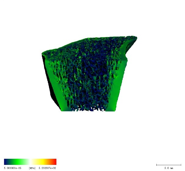 FEA can be used to calculate bone stiffness, failure load and more. |
Cortical density/thickness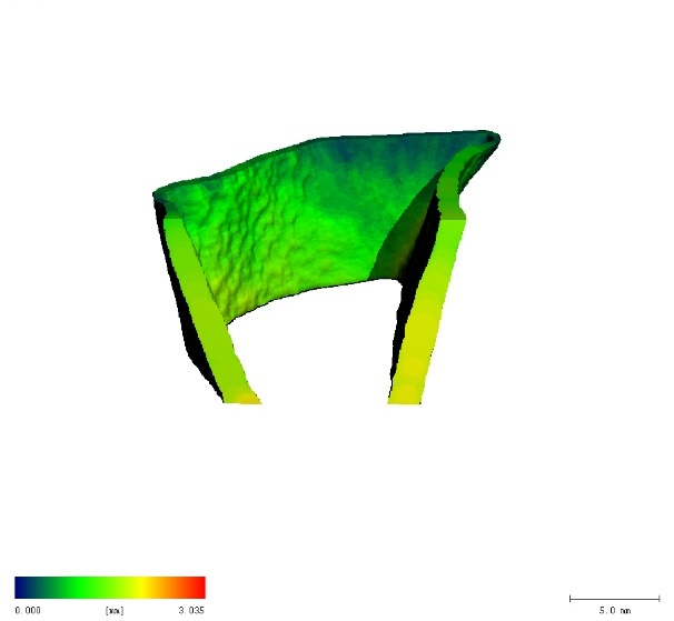 | Trabecular density/thickness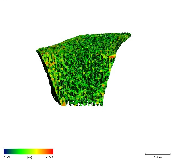 |
| Taking bone micro-architecture into account in combination with bone density improves fracture risk prediction and may aid in the management of patients with osteoporosis. This imaging modality uses a low dose of x-ray radiation and is otherwise non-invasive. You can read more about our device here. | |