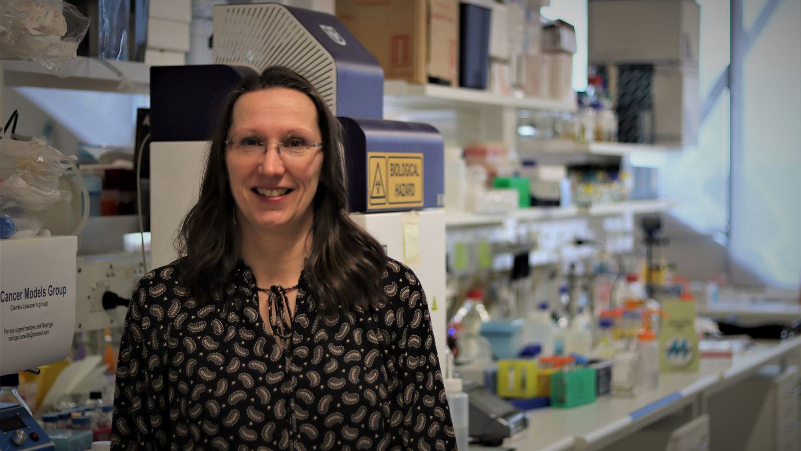
Daniela Loessner
Tissue-engineered 3D models help unlock secrets of cancer
For decades, scientists have been growing cancer cells in the laboratory to study their characteristics, behaviour, and responses to treatments in a controlled environment.
But, contrary to long-held perceptions, cancer isn’t simply a chaotic mass of aberrant cells growing and multiplying out of control.
More and more research shows that cancer cells are highly influenced by, and in turn influence, the environment in which they grow, leading to the concept of a tumour as a complex, structured and interconnected ‘organ’.
That's where engineering is increasingly playing a role in cancer research.
“It’s become clear that we can’t meaningfully model the biology of tumours in the laboratory without engineering input,” explains Associate Professor Daniela Loessner.
“To better understand cancer, we need models that mimic the 3D structure and function of tumours and even the mechanical forces that shape their dynamics.”
Associate Professor Loessner holds joint appointments in the Faculty of Engineering and the Faculty of Medicine, Nursing and Health Sciences at Monash University, and also at the Leibniz Institute of Polymer Research in Dresden, Germany.
An international expert on tissue engineering in cancer, Associate Professor Loessner and her research colleagues were invited to author a comprehensive review of the field for the prestigious international journal Nature Reviews Materials, published in May 2023.
“Cell biologists can grow cancer cells in the laboratory but we can’t understand tumours by growing those cells in isolation,” she argues. “The secret to designing better 3D cancer models is multidisciplinary collaboration between bioengineering and biology.”
While engineers are familiar with biomaterials and their properties, cancer biologists know how cells grow and reproduce and what sort of analyses need to be performed to understand disease progression and responses to treatments.
Advanced tissue-engineered 3D models that accurately resemble the “tumour microenvironment” - the niche in which a tumour develops - can greatly accelerate discovery and clinical translation by closely replicating physiological conditions.
They are also becoming powerful platforms for evaluating the efficacy and safety of new therapies and combinations of therapies, and even for high-throughput drug screening and discovery.
The tumour microenvironment contains not only cancer cells but also various other types of non-cancerous or “stromal” cells, as well as components of the “extracellular matrix”, all of which are subject to mechanical stimuli.
Biomechanical characteristics can also affect oxygen and nutrient levels at different locations within the tumour.
In their laboratories, Associate Professor Loessner’s teams at Monash and Leibniz build the matrix of the tumour - the scaffold and material within which the tumour grows - from composite biomaterials developed by combining natural and synthetic polymer building blocks.
Advanced techniques including additive manufacturing (“3D printing”) using polymers and bioprinting - in which droplets or strands of biomaterial “inks” are fired through microscopic nozzles - have significantly improved the ability of engineers to build biologically relevant scaffolds.
Once the scaffolding is in place, cancer cells are introduced, together with different stromal cells that surround them.
Much of the behaviour of tumours and even responses to treatments are now being linked to the crosstalk between cancer cells and stromal cells. Indeed, the healthy stromal cells that support the tumour’s growth may themselves be a target for anti-cancer therapies.
Cancer cells can also secrete and remodel their own extracellular matrix, changing the biomechanical properties of surrounding tissues in a way that may enhance cell invasion.
“For example, pancreatic tumours are as hard as a walnut,” explains Associate Professor Loessner, “and that hardness can be associated with more aggressive disease that’s much more difficult to treat.”
Consequently, Associate Professor Loessner and colleagues are also finding and implementing ways to make the stiffness and elasticity of the 3D models “tunable” so they can mimic the properties shown by tumour tissues at different stages of disease progression.
"By replicating the biomechanical properties and the physical environment, we can get tumour cells to grow in the laboratory just as they would in their original setting in the body,” she says.
The better the tissue-engineered 3D model, the more closely researchers will be able to study and understand cancer cell growth, proliferation, migration and invasion, and observe responses to chemotherapies and the rapidly-developing range of immunotherapies or stromal-targeting therapies.
Ultimately, says Associate Professor Loessner, 3D tumour engineering also raises fascinating possibilities like “co-clinical trials” in which the patient and laboratory models of their tumour receive different treatments and the effects can be directly compared.
“This would enable us to test different drug combinations and find the ones that will benefit the patient much more rapidly.”